In hospitalized elderly, pediatric, or unconscious patients, a common issue to address is the transfer of gases or drug substances into the body orally or via the nasal cavity. To solve the purpose, endotracheal tubes, abbreviated as ETT, can be employed. The proper administration of gases and medications via ETT ensures the delivery of the substances into the airway passage.
Structure of ETT
An ETT is based on tubing with varying lumen sizes. It provides the space for the air or drugs to pass into the body when administered from its adaptor or connector. ETT can be prepared by using the following materials:
– Polyvinyl chloride (PVC)
– Nylon
– Silicone rubber
– Teflon
– Latex rubber
– Stainless steel
Among these, PVC is the preferred material due to its stability and flexibility at room temperature. It does not hinder the visibility of the exhaled air i.e. breath fogging due to its clarity thus enabling the physician to visualize the contents.

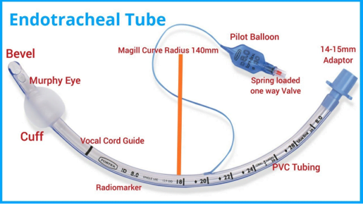
Parts of an endotracheal (ET) tube
The inflatable cuff
In addition to the tube, cuffs are attached to its one end. Cuffs are the balloons that, once into the trachea, are inflated to provide resistance with the tracheal wall which hinders the tube from displacement. Moreover, the tube creates a positive pressure inside the airway passage to aid ventilation. The discharge of gastric contents into the lungs is also prevented by the cuff of ETT.

Inflating process of the cuff of the ETT
The bevel
Bevel is the angled or slant end of ETT which penetrates deep into the respiratory passage. It helps in the proper placement of the tube without damaging the vocal cords. The visualization of the endotracheal pipe is enhanced by the bevel.
The Murphy’s eye
The Murphy’s eye is the opening near the bevel which allows airflow in case of the blockage of the bevel. In this condition, Murphy’s eye provides ventilation thus preventing the complete obstruction of the airflow.
The connector/adaptor
The adaptor or connector is the connection point between the tube and the mechanical ventilator tubing. Its diameter is 14 to 15 mm in general which is suitable for both adult as well as pediatric patients.
Sizes of ETT
ETT has an internal diameter of the lumen falling between the range of 2.0 to 12.0 mm among which 7.0 to 7.5 mm tubes are suitable for female patients while 7.5 to 8.5 mm are appropriate for male ones. A narrow lumen indicates greater airflow resistance.
Types of ETT
Endotracheal tubes can be classified according to their delivery via the oral cavity i.e. oropharyngeal tubes or nasal cavity i.e. nasopharyngeal tubes. These are available in different sizes, beveled or non-beveled, cuffed or non-cuffed. Some of the types of endotracheal tubes are as follows:
– RAE PVC or wire-reinforced silicone rubber tubes: RAE tubes have been named after its inventors i.e. Ring, Adair and Elwyn. They are oral tubes with a pre-formed bend to minimize the risk of tracheal obstruction. Oral RAE tubes can be further divided into south-facing and north-facing tubes based on their placement in the oral cavity. If the tube connector is facing the patient’s feet, the tube is termed as south-facing while the north-facing tube indicates a direction towards the patient’s head.

RAE ET tube with its pre-formed bend
– Bivona Fome-Cuff tubes: Bivona Fome-Cuff tubes are useful during laser surgery of the respiratory tract.
– LITA ETT: LITA endotracheal tubes are employed to deliver local anesthesia into the larynx and trachea.
– PneuX ETT: Pneux endotracheal tubes are also called Young Lo-Trach tubes. These are rigid, straight, and flexible silicone-based tubes. Pneux tubes are known to cause lesser endotracheal traumas.
– Double-lumen tubes (DLT): Double-lumen tubes (DLT) such as Carlens, White, and Robertshaw tubes are employed during lung or thoracic surgeries. These can be left or right-sided.

Double-lumen tubes: (a) right-sided and (b) left-sided
Placement of an ETT
The ETT tube is placed into the trachea in the following steps:
– Firstly, it is ensured that the patient is lying down straight. The patient, if conscious, is asked to stay calm during the process.
– The ETT is taken out from its packaging and checked for its intactness before its administration.
– The distal end of the ETT bearing the bevel is introduced into the trachea and is gently pushed deeper into the airway passage. If the patient feels uncomfortable, the process should be stopped for a while until the patient is relaxed and then the tube can be inserted again.

An ETT placed into the airway passage
– Once the tube is placed, the cuff is inflated using normal saline (0.9% NaCl aqueous solution) in order to fix the tube in its place.
– The adaptor or connector is attached to the mechanical ventilation machine for the purpose of administering oxygen.
Applications of ETT
The ET tubes provide the following advantages:
– Maintenance of airway patency
– Aids the respiratory ventilation
– Suctioning of gastric or oropharyngeal secretions
– Anesthetic delivery via airway passage i.e. administration of desflurane, isoflurane, sevoflurane, etc.
– Drug delivery (i.e. salbutamol, atropine, epinephrine, ipratropium, lidocaine, etc.) in case of emergency or among bedridden/elderly patients
Demerits of the ETT
Although ETT is safe to use, their placement, if not done properly, may result in the following:
– Obstruction of the airway passage may result in apnea i.e. breathing cessation.
– Oropharyngeal trauma if the tube does not fit properly.
Contraindications of ETT
The ETT is contraindicated in patients with:
– Expanding neck hematoma i.e. presence of coagulated blood in the tract
– Active epistaxis i.e. nasal bleeding
– Head trauma Facial trauma
Conclusion
Delivery of oxygen, as well as drugs, is a major issue in hospitalized patients, elderly subjects, pediatrics as well as unconscious patients. ETT is an easy-to-use and inexpensive means to solve the problem. Their placement is easy and quick and poses no serious side effects in the patients after removal. Thus, the ETT is one of the most commonly employed and preferred medical instruments by physicians.

PhD Scholar (Pharmaceutics), MPhil (Pharmaceutics), Pharm D, B. Sc.
Uzma Zafar is a dedicated and highly motivated pharmaceutical professional currently pursuing her PhD in Pharmaceutics at the Punjab University College of Pharmacy, University of the Punjab. With a comprehensive academic and research background, Uzma has consistently excelled in her studies, securing first division throughout her educational journey.
Uzma’s passion for the pharmaceutical field is evident from her active engagement during her Doctor of Pharmacy (Pharm.D) program, where she not only mastered industrial techniques and clinical case studies but also delved into marketing strategies and management skills.
Throughout her career, Uzma has actively contributed to the pharmaceutical sciences, with specific research on suspension formulation and Hepatitis C risk factors and side effects. Additionally, Uzma has lent her expertise to review and fact-check articles for the Health Supply 770 blog, ensuring the accuracy and reliability of the information presented.
As she continues her PhD, expected to complete in 2025, Uzma is eager to contribute further to the field by combining her deep knowledge of pharmaceutics with real-world applications to meet global professional standards and challenges.