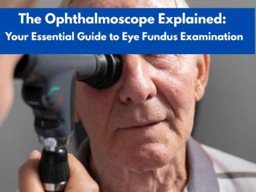
The Ophthalmoscope Explained: Your Essential Guide to Eye Fundus Examination

According to the National Library of Medicine, nearly 10 million people in the US are suffering from diabetic retinopathy, a condition associated with retinal damage due to high blood sugar levels. The condition can be diagnosed using an ophthalmoscope that visualizes the internal structures of the eyes, making it easy for the ophthalmologist to diagnose a disease state, if present.
Proper eye examination helps detect diseases at an earlier stage
In this article, we will go through the basic introduction of ophthalmoscopes and learn how they are useful for eye fundus examination. In addition, some of the well-known ophthalmoscope brands have also been linked for the reader’s convenience.
What is Eye Fundus Examination or Ophthalmoscopy?
The term fundus of the eye is used to refer to the back surface of the human eye that includes the retina (third layer of the eye), macula, optic disc, fovea, and blood vessels in the area. As the retina contains rod cells and cone cells (photoreceptor cells), this area is associated with vision.
A fundus examination is the visualization of the eye fundus using an ophthalmoscope. The purpose is to evaluate the condition of the optic nerve and blood vessels in terms of the level of damage occurred to them. In addition, ophthalmoscopy also helps diagnose the presence of retinal detachment. This fundus examination becomes particularly essential in conditions like glaucoma, macular degeneration, diabetic retinopathy, etc.
What are Ophthalmoscopes?
An ophthalmoscope is a handheld tool designed to examine the internal structure of the eyes, the process of which is called ophthalmoscopy or fundoscopy. As mentioned above, it helps visualize the retina where the image is formed due to the presence of photoreceptor cells.
In addition, the optic nerve can also be analyzed, that takes information from the retina to the brain. Ophthalmoscopy also enables the ophthalmologist or optometrist to check the condition of blood vessels.
Parts of an Ophthalmoscope
Before diving into the working of an ophthalmoscope, let us first learn about its parts. As an ophthalmoscope has a head and a handle, the following section covers the components of both:
Ophthalmoscope Head
Light Source
A light source is an electric bulb attached to the device that provides the necessary illumination to visualize the internal structure of the eye.
Mirror or Prism
A mirror or prism in an ophthalmoscope reflects the light coming from the bulb at a right angle into the patient’s eye through the viewing window.
Lens Wheel
The lens wheel or diopter dial helps the user to rotate a series of lenses, all of which have different powers. This way, a clear and focused image of the internal structures of the eye can be achieved which ensures the accuracy of the procedure.
Viewing Window
The viewing window is the component of the ophthalmoscope through which the user looks into the interior parts of the eye.
Aperture Dial
The aperture dial of an ophthalmoscope allows the healthcare provider to alter the size of the light beam. This is important because different sizes of the light beams are used for different purposes.
Filter Switch
A filter switch allows the user to choose lights with different colors to make the examination clearer. For instance, a red-free light is used for examining the blood vessels in depth, or a cobalt blue light is selected for use with fluorescein dye.
Ophthalmoscope Handle
Power Source
The rechargeable or disposable energy source (batteries) come with an ophthalmoscope to give the necessary power to its light bulb.
On/Off Switch or Rheostat
An on/off switch or rheostat in an ophthalmoscope helps control the power of the device. This way, it allows the adjustment of the brightness of the light source depending on the patient’s needs.
Eyebrow Rest
Eyebrow rests are added in the design for the user’s convenience.

Most ophthalmological examinations only take 10 to 15 minutes to conduct
How Does an Ophthalmoscope Work?
In simpler terms, an ophthalmoscope works by illuminating the interior of the eye with the help of a mirror and a light source. In the first step, a light beam from the light source or electric bulb enters the eye and reaches the retina after crossing through a tilted mirror and then a lens.
Next, it is reflected back and reaches into the observer’s eye through a peephole at the center of the mirror. As a result, a magnified image of the retina is formed that is virtual and erect. To correct the refractive optical errors in the process present due to issues like myopia or hypermetropia in both the patient and the ophthalmologist, a series of concave and convex lenses is present in the system that can be rotated to achieve clear visuals.
Frequently Asked Questions
The following section covers some of the frequently asked questions by patients regarding the ophthalmoscopy exam:
1. How long does an ophthalmoscopy examination take?
An eye examination done using an ophthalmoscope usually takes 5 to 10 minutes. However, your ophthalmologist might ask you to come earlier if the pupil-dilating drops are to be used prior to the examination. These drops dilate the pupil and, therefore, make it easy for the doctor to look into the internal structures of the eye. Once used, these drops may require 15 to 30 minutes to cause pupil dilation, thus adding to the overall time of fundoscopy.
2. How to prepare for an ophthalmoscopy?
Before performing an ophthalmoscopic procedure, there may be a need to use eye drops for pupil dilation. The purpose is to expand the pupil so that the retina and other parts of the fundus can be seen clearly during the examination. As these eye drops make the vision blurred for a few hours, it is better not to drive. Also, if your job requires the use of heavy machinery or anything that needs you to see clearly, it is also advised to take a day off.
Additionally, if you have any allergies to certain medications, let your doctor know about them. This will also help her decide which medicines should be administered to you before, during, and after the procedure.
There is also a risk of drug interactions, as some of the medications (both the over-the-counter and prescription medications) you already take may interact with the eye drops. Similarly, certain dietary supplements may also restrict their dilation effect. In such cases, your doctor might ask you to discontinue these products for a few days before the procedure.
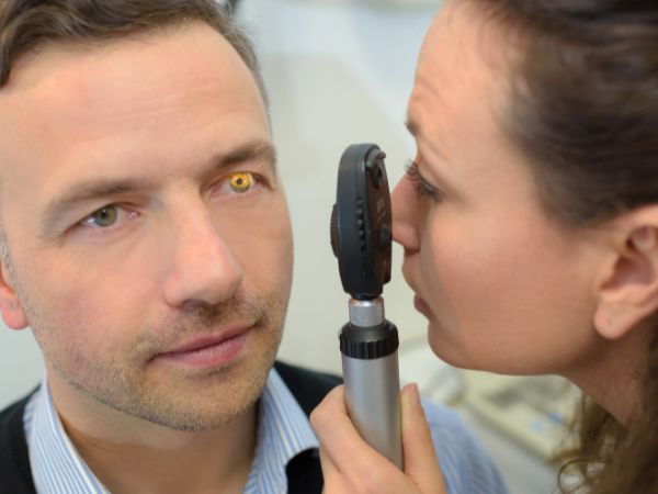
3. Does an ophthalmoscopy exam hurt?
No, an ophthalmoscopy examination is not painful. Rather, it is a quick way to check the internal structures of the eyes. However, the patients may feel mild discomfort due to the bright light or the clicking sound from the instrument when the doctor adjusts the device. Keep in mind that the pupil-dilating drops do cause a stinging sensation for a few hours.
4. How are the results of an ophthalmoscopy examination interpreted?
If the ophthalmologist sees a normal retina, optic disc, and blood vessels, the internal structures of the eyes are healthy. However, if the retina is swollen or has spots on it, this is an indication of a disease state. The issue might be one of the following and is to be confirmed by the ophthalmologist:
- Diabetes
- Damaged optic nerve
- Melanoma or cancer affecting the eyes
- Cytomegalovirus (CMV) retinitis, which is an infection affecting the retina
- High blood pressure or hypertension
- Glaucoma
- Macular degeneration or loss of sharp vision due to aging
- Retinal tear/detachment or separation of the retina from the back of the eye
5. Are there any risks associated with an ophthalmoscopy examination?
Ophthalmoscopy is a safe procedure that does not pose any risk to the patients. However, some people might feel uncomfortable during and after the procedure and report some of the following problems, which are often associated with the use of eye drops for pupil dilation:
- Dry mouth
- Dizziness
- Flushing
- Nausea
- Vomiting
- Narrow-angle glaucoma
If these symptoms persist after a few days of treatment, consult your doctor.

Ophthalmoscopy is a painless process that increases patient comfort during eye examination
Ophthalmoscopes and Related Parts at Health Supply 770: Features and Specifications
An ophthalmoscopy is a process of visualizing the internal structures of the eyes. For this purpose, ophthalmoscopes have been designed that shine a bright light into the eye to get a clear image. The doctor is then able to tell whether the internal structures of the eye are healthy or not, depending on the presence or absence of the signs of any damage.
However, when it comes to learning about ophthalmoscopes, it is also essential to look into some of the products that many brands manufacture. Some of these well-known ophthalmoscopes and their related parts are also available at Health Supply 770 and have been detailed below, along with their features and purchase links:
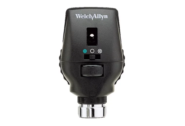
Welch Allyn Coaxial Ophthalmoscope for Eye Examination– 3.5V LED 11720-L
US$264.88
Product Details
Welch Allyn coaxial ophthalmoscope is a handheld tool that is employed for the examination of the eye fundus. It helps the healthcare provider to look into the internal structures of the eye and diagnose the signs associated with an eye disease with maximum clarity. As the device allows a shadow-free viewing of the eye fundus, the image formed in the eyes of the observer is clear and free from the disturbances caused due to glare. Resultantly, an in-depth eye examination becomes possible.
In addition, these Welch Allyn coaxial ophthalmoscopes also come with a 3.5V LED illumination that lasts for longer and, therefore, eliminates the need of frequent battery change. To meet the varying diagnostic needs, the device has 18 unique aperture or filter combinations. This ophthalmoscope head can be employed with all other Welch Allyn equipment.
Welch Allyn 03000U6 3.5V Halogen Lamps, Ophthalmoscope for 11710/8000, BX6

https://hs770.com/product/welch-allyn-halogen-bulb-for-ophthalmoscope/
Product Details
Welch Allyn 03000U6 3.5V halogen lamp for ophthalmoscopes is an item that comes with a bi-pin base, indicating the presence of two small pins on the product that are needed for its attachment with the ophthalmoscopic device. The device acts as a source of light and, therefore, helps in the visualization of the internal parts of the eye, i.e., the eye fundus.
Moreover, these Welch Allyn 03000U6 3.5V halogen lamp for ophthalmoscopes are long-lasting as compared to the incandescent bulbs, as the manufacturer claims an average lifespan of about 2000 hours. Furthermore, the halogen bulbs employed in these ophthalmoscopy lamps are usually single or double-coiled.
*Note: The prices mentioned in the article are taken from the Health Supply 770 website. These may vary depending on the vendor.

Ophthalmoscopes help identify several eye diseases within minutes
The products linked in the article, while learning about ophthalmoscopes, along with many other medical supplies, can be purchased from Health Supply 770, a reliable name when it comes to medical products. They have a 30-day money-back guarantee and provide your products to you in the shortest possible time. Click the link at the end of the article to check the wide range of eye examination products.
Conclusion
Eyes are one of the most sensitive parts of the human body. As they are constantly exposed to the environment, the risk of damage to the internal structures is quite high. Therefore, a regular examination of the eye fundus, known as ophthalmoscopy, is recommended.
In this regard, an especially designed instrument called an ophthalmoscope is used to visualize the internal structures of the eyes. The device works by shining a bright light on the retina, optic nerve, fovea, and blood vessels, which is then reflected and reaches the eye of the doctor. Resultantly, a clear image is created that helps the visualization of the signs of any disease. Overall, the method is quick and easy, in addition to being fully pain-free.
Considering their importance, ophthalmoscopes are an integral part of eye examination and are, therefore, needed within all the relevant healthcare facilities. For purchasing different kinds of medical devices, including ophthalmoscopes, reliable vendors like Health Supply 770 should be approached. They ensure the provision of quality products along with satisfactory services.
References
https://pmc.ncbi.nlm.nih.gov/articles/PMC10273133/
Read also
Otoscope and Ophthalmoscope: Key Differences and Uses Explained