Benefits of using Evans Elite Endoscopic Instrument Set
Endoscopy is a painless medical imaging and sampling technique that enables the physicians to look inside the patient’s body with the help of a minute camera called an endoscope which is fixed on a long tube and is passed down the patient’s gastrointestinal cavity via the throat. It takes 10 to 15 minutes to complete an endoscopic evaluation.
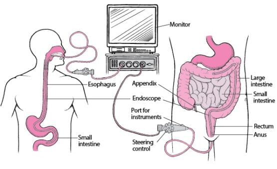
An upper and lower endoscopic evaluation
Why is an endoscopy done?
Endoscopy is done for various reasons which include:
• Tissue sampling (biopsy)
• Evaluation of inflamed areas
• Detection of cancer
• Detection of ulceration or other celiac diseases
• Detection of gastroesophageal reflux disease
• Structural analysis of the digestive tract
• Detection of blockage points in the alimentary canal
• Treatment of a gastrointestinal polyp
• Widening of the esophagus
• Burning of a bleeding vessel
• Detection of the causes of diseases in case the symptoms of nausea, heartburn, vomiting, abdominal pain or gastrointestinal bleeding do not subside after treatment
Parts of an endoscope
An endoscope is a medical instrument which is a combination fiber-optics technology and charge-couple devices (CCD). Its diameter ranges between 2 to 6 mm depending upon the use. In an endoscopic instrument set, the fiber-optics illuminates the body viscera and the charge-couple device captures an image which is then illustrated on a connected screen. It mainly consists of an insertion tube with a tip. A control section provides the user authority over the device while in use. The following image illustrates an Evans Elite endoscopic instrument set with its parts labeled.
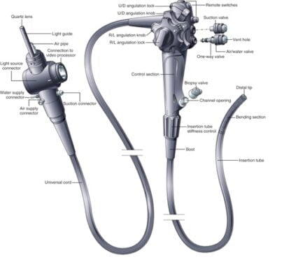
An endoscope with labelled parts (Evans Elite Endoscopic Instrument Set)
An Evans Elite Endoscopic takes pictures of the internal body parts in monochrome and reproduces them to generate a final colored image. With latest high tech endoscopes, it is also possible to zoom in the images and magnify the diseased areas.
Types of endoscopies
As endoscopy simply means visualizing the internal body parts, the endoscopic approaches can be divided into the following types depending on the area of the body under examination.
- Esophagogastroduodenoscopy: Esophagogastroduodenoscopy is the endoscopic evaluation of the esophagus, stomach, and duodenum. It is helpful for visualizing the digestive tract and highlighting the affected areas.
- Enteroscopy: Enteroscopy is the endoscopic assessment of the parts of the small intestine.
- Sigmoidoscopy or Colonoscopy: Colonoscopy is performed to evaluate the colon, the second segment of the large intestine. The aim is to look for the causative agents of the conditions like chronic diarrhea or unexplained rectal bleeding. Additionally, the affected tissues in diseases such as colon cancer can also be imaged and sampled.
- Rectoscopy: Rectoscopy is the endoscopic assessment of the rectum, the last part of the large intestine.
- Anoscopy: Anoscopy is the endoscopic evaluation of the anal area. It helps in the visualization of tearing of torn parts of the anal tissue, dysplasia (abnormal cellular growth) as well as cancer tissues.
- Otoscopy: Otoscopy endoscopically evaluates the internal parts of the ear including tympanic membrane, external auditory canal as well as the parts of the middle ear.
- Cystoscopy: Cystoscopy is the endoscopic evaluation of segments of the urinary tract including urinary bladder and urethra.
- Rhinoscopy: Rhinoscopy, also called nanoscopy, is the endoscopic assessment of the nasal cavity.
- Bronchoscopy: It is the endoscopic evaluation of the lower respiratory system.
- Colposcopy: Colposcopy is done to closely check the internal structure of the cervix. The changes in the cervix caused by human papillomavirus (HPV) or cervical cancer can easily be seen by this technique.
- Hysteroscopy: Hysteroscopy is the endoscopic evaluation of the uterus in case the symptoms such as unexplained bleeding, pelvic and vaginal pain, or menstrual difficulties appear.
- Falloposcopy: Falloposcopy is the endoscopic assessment of fallopian tubes. Along with this, the technique can also help in the removal of cellular debris from the tube.
- Laproscopy: Laproscopy is the endoscopic evaluation of the abdominal cavity.
- Arthroscopy: Arthroscopy is employed for the endoscopic imaging of joints with minimum invasion.
- Thoracoscopy: Thoracoscopy is the endoscopic assessment of the chest area. It is recommended for the imaging and sampling of the tissue in case of lung cancer as well as in other diseases.
Preparing for an endoscopy
• The patient must be in a fasting state for endoscopic surgeries.
• Eating should be stopped at 8 hours and drinking should be stopped at least 4 hours prior to the test. Medications can be taken if necessary as guided by the physician.
• However, blood thinning medications need to be stopped as they may elevate the chances of bleeding during the procedure.
• Laxatives may be prescribed to the patient to clear out any waste material out of the alimentary canal.
An endoscopic evaluation
• The patient is often recommended intravenous sedatives just before an endoscopic surgery to relax them.
• Breathing rate, heart rate, and blood pressure are checked.
• A plastic mouth guard is placed into the patient’s mouth to keep it open for longer.
• Air can be fed into the patient’s gastrointestinal tract to inflate it and make it easier for the endoscope to pass through it.
• An anesthesia is sprayed into the oral cavity to numb the tissues and ease the transfer of the endoscopic camera attached to a tube.
• Finally, the endoscopic instrument is inserted into the esophagus.
• As soon as the camera starts showing the patient’s insides, the physician looks for signs of disease.
• Images and tissue samples may be taken during the procedure for further use.
After the endoscopic surgery
• The patient may feel dizzy due to the anesthesia so driving and working should be avoided for some time. It is advised to take rest at least for a day until the sedative effect wears off.
• It is normal to feel bloating. This is due to the gas pumped into the body while conducting the procedure.
• Muscle cramps can also be experienced in the examined area.
• Sore and painful throat is also a common thing after an endoscopic evaluation.
• The results of the endoscopy are given to the patient as soon as the test is completed. However, in the case of a biopsy, the results may take a few days to be ready.
• If the pain does not subside after some hours, it is advised to contact your healthcare practitioner for further assistance.
Can endoscopic surgery be harmful?
Endoscopic approaches, if not done the right way, can cause some issues in the patient’s body. These include the following:
- Gastrointestinal bleeding: In the case of tissue sampling called a biopsy, a small piece of the gastrointestinal tissue is removed and tested in the lab to find out the presence of possible diseases. This may, sometimes, result in bleeding at the area from where the tissue has been excised. This bleeding is rarely critical enough to need a blood transfusion.
- Damage of gastrointestinal tract: In some cases, gastrointestinal tract can get a tear by the endoscopic surgical instruments while conducting an endoscopy. Mostly, the damage happens in the esophagus but any other part can also be affected. If severe, this may need hospitalization followed by surgery.
- Infections: Although the risk is low an endoscopic instrument set can potentially transfer microbes into a person’s digestive tract resulting in an infection. Thus an antibiotic course may be required to get rid of the infection. Physicians sometimes recommend antibiotics as a prophylactic treatment i.e. to prevent the patient for a possible future infection.
Conclusion
Endoscopy is a safe and reliable method for the visualization as well as a sampling of the internal body parts. It is a minimally invasive technique that is practically painless. As most of the surgical and medical instruments and procedures come with some discomfort, endoscopic surgery can also cause pain and minor side effects afterward but overall, it is a risk-free method to adopt when an internal medical checkup is needed.
