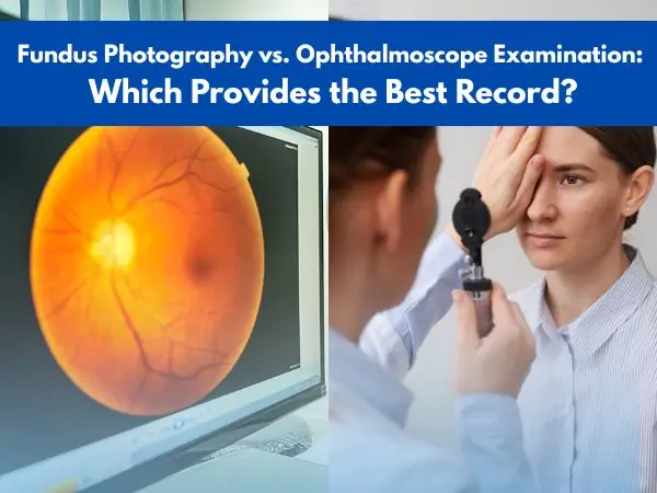
Fundus Photography vs. Ophthalmoscope Examination: Which Provides the Best Record?
According to the Centers for Disease Control and Prevention (CDC), approximately 7 million Americans suffer from vision impairment. In most of these cases, healthcare providers recommend a regular fundus examination that can be either conducted by fundus photography or an ophthalmoscope examination. But what are these two, and how do they differ?
A timely eye examination helps diagnose diseases at their earlier stages
In this article, we will go through the differences between fundus photography and ophthalmoscope examination to learn which of these can provide better results. In addition, some of the well-known ophthalmoscope brands have also been linked for the reader’s convenience.
What is Ophthalmoscope Examination?
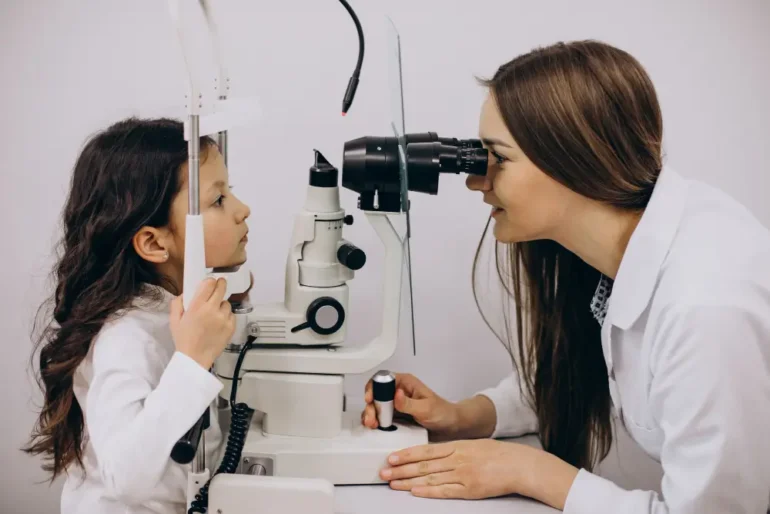
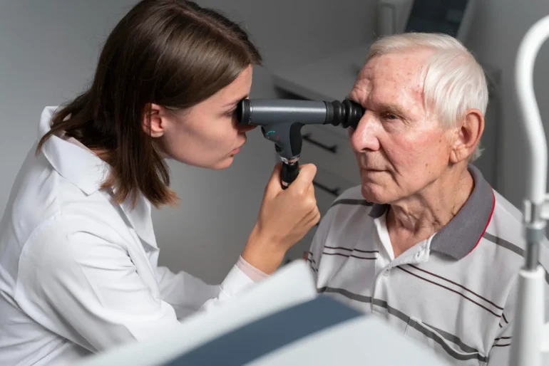
An ophthalmoscope examination, also known as ophthalmoscopy or fundoscopy, is a method of visualizing the internal parts of the eye, primarily the fundus of the eye, i.e., retina (third layer of the eye), macula, optic disc, fovea, and blood vessels in the area. It is done by using a handheld ophthalmoscope that sends a beam of light in the eye, followed by receiving it and creating an image for the examiner.
Ophthalmologists or optometrists use this method for the diagnosis of several eye-related conditions, like glaucoma, macular degeneration, diabetic retinopathy, etc. As an ophthalmoscope examination does not take much time, it can be easily completed within 10 to 15 minutes.
Advantages
When it comes to conducting an ophthalmoscope examination, the healthcare provider aims to achieve some of the following benefits from the technique:
Magnified View of the Eye Fundus
As an ophthalmoscope can magnify the view of the central retina, i.e., up to 15X, it can provide a clearer image of the internal parts, which eases the diagnosis. The image produced is also upright and unreversed.
Portability
An ophthalmoscope examination is easy to conduct due to the ease of usage of the instrument known as an ophthalmoscope. As ophthalmoscopes are small-sized, handheld devices, they can be easily carried around and used in different clinical setups.
No Need for Pupil Dilation
As most eye procedures require the use of pupil-dilating drops, an ophthalmoscope examination can be conducted without them. However, in some cases, there may be a requirement if the procedure demands.
Easy to Use
Ophthalmoscopes are easy to use instruments and can be mastered by medical students and general practitioners within a few attempts. To further ease the learning process, a 7-step protocol is also available that has been covered in another article.
Disadvantages
Despite the fact that an ophthalmoscope exam is the easiest method of conducting a fundus examination, the method is also associated with certain disadvantages, some of which have been given as follows:
Small Field of View
An ophthalmoscope provides a smaller field of view that often ranges from 10 to 12 degrees. This indicates that the examiner can only see a small portion of the retina at a time. This keyhole vision limits the efficacy of the procedure as it may take more time to complete the fundus examination.
Lack of Stereoscopic Vision
The lack of stereoscopic vision during an ophthalmoscope examination is a limitation of the instrument rather than the examiner or the method. This is because direct ophthalmoscopes are not designed to perceive the depth of the retina by generating a 3D image. However, indirect ophthalmoscopes have solved this issue as they come with prisms and mirrors that make 3D viewing possible.
Low Patient Comfort
As an ophthalmoscope examination is conducted by directly looking into the patient’s eye, the patient needs to be upright and alert during the procedure. Moreover, a shorter distance needs to be maintained between the device and the patient’s eye, which often makes people uncomfortable.
Eye-related diseases are extremely prevalent in the US, especially among diabetic and hypertensive patients
What is Fundus Photography?
Fundus photography is a specialized eye examination technique that involves the use of a high-resolution camera so that clear and highly accurate images of the eye fundus can be captured. The method is useful for the diagnosis, monitoring, and management of eye diseases, such as diabetic retinopathy, glaucoma, macular degeneration, etc.
To conduct fundus photography, a beam of light is used to illuminate the eye fundus, and a picture is clicked through the pupil. As the images taken this way are digital, they can be stored for future analysis as well.
Advantages
As fundus photography is a newer method compared to taking an ophthalmoscope examination, it provides numerous benefits, some of which are as follows:
Non-Invasive Method
Taking a fundus examination is a quick and easy process that is often preferred by patients due to its non-invasive nature. The procedure is also painless and does not cause any major side effects, apart from temporary light sensitivity often associated with the use of pupil-dilating drops before the test.
Wide-Field Views
The product also ensures the provision of a wider field of view as it is capable of capturing a larger retinal area as compared to that covered during a traditional ophthalmoscope examination.
Permanent Visual Record
The photographs taken during this procedure are digital, permanent, and have a high resolution. This way, the image clarity is enhanced. Moreover, these images can be saved in the database for future analyses and, therefore, come in handy for checking the disease progression as well as the efficacy of the treatment. As these images are also shareable, these can be transferred from one doctor to another during patient referrals.
Quick Disease Detection
Fundus photography, when compared to an ophthalmoscope examination, is far superior at spotting early signs of numerous eye diseases, including macular degeneration, diabetic retinopathy, hemorrhages, and glaucoma.
Higher Patient Compliance
As the fundus photography just requires the patient to sit upright for a few minutes, the procedure is well-tolerated, especially by those who are taking it for the first time.
Cost-Effectiveness
The method is cost-effective as compared to the ophthalmoscope examination, as the need of a trained specialist is eliminated. In addition, the equipment used for fundus photography is also cheaper.
Disadvantages
Despite its several advantages, fundus photography also comes with certain disadvantages that include the following:
2-Dimentional Images
The images taken by fundus photography are 2D and, therefore, do not allow the estimation of depth, thus restricting diagnosis in some cases.
Poor Image Quality
In some cases, the pictures taken during fundus examination are not as clear as necessary for the analysis of the condition, leading to a compromised diagnosis.
Expert Dependency
Although an eye specialist is not needed to conduct the procedure, it is still necessary that a skilled technician takes the images of the eye fundus. This way, both the clarity and the accuracy of the images are ensured.
Mobility Issues
As compared to handheld instruments, like ophthalmoscopes, the instruments used for fundus photography are bigger and bulkier and, therefore, not mobile.
Fundus Photography vs. Ophthalmoscope Examination: Which One is Better?
When it comes to a comparison between the fundus photography and the ophthalmoscope examination, the former one is considered highly accurate and reliable. This claim is supported by a study published in the Neuro-Ophthalmology Journal in which 587 patients were recruited for the examination of their eye fundus in the emergency department.
The results of the study have clearly indicated the superiority of fundus photography over the ophthalmoscope examination, as higher image quality is believed to ease the identification and diagnosis of abnormalities as well as diseases of the eyes.

Pupil-dilating drops are used before fundus examination to ease the process
Ophthalmoscopes and Related Parts at Health Supply 770: Features and Specifications
An ophthalmoscope examination is a method of looking into the eye fundus with the aim of diagnosing possible abnormalities or diseases. The method is simple and does not consume much time, a feature rendering it preferable by both the patients and the healthcare providers.
However, when it comes to learning about ophthalmoscope examination and fundus photography, it is also essential to look into some of the products that many brands manufacture. Some of these well-known ophthalmoscopes and their related parts are also available at Health Supply 770 and have been detailed below, along with their features and purchase links:
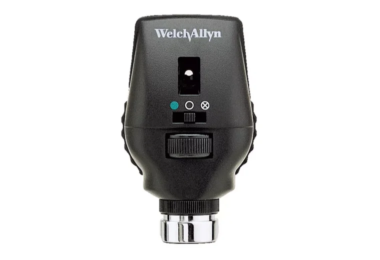
Welch Allyn Coaxial Ophthalmoscope for Eye Examination– 3.5V LED 11720-L
US$264.88
Product Details
Welch Allyn coaxial ophthalmoscope is a handheld tool that is used for the examination of the human eye and make a diagnosis of the diseases affecting the fundus. The device is known for providing high-quality visuals of the internal parts of the eye and, therefore, ensures image clarity. Due to its shadow-free as well as glare-free viewing, the view does not become blurred or distorted. As a result, an in-depth examination of the eye is possible.
In addition, the light source of these Welch Allyn coaxial ophthalmoscopes is a 3.5V LED, a product known for providing long-lasting illumination, thus reducing the need for frequent battery recharging. The device can also be employed with all kinds of relevant Welch Allyn equipment. Furthermore, the 18 unique apertures or filter combinations also come with the device to meet the varying diagnostic needs.
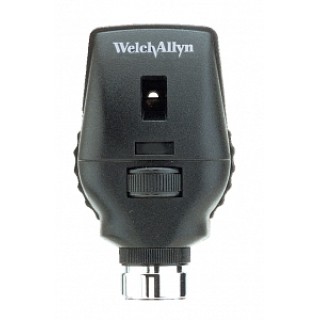
Welch Allyn 11710 Halogen Ophthalmoscope Head – 3.5V
US$182.88
Product Details
Welch Allyn 11710 halogen ophthalmoscope head is a high-performance eye-examination tool that has been designed for eye examination in a precise and reliable manner. The product is known for the bright light it delivers to the eye fundus to cause its illumination, resulting in the visualization of the retina, its blood vessels, optic disc, etc.
Moreover, these Welch Allyn 11710 halogen ophthalmoscope heads are highly durable instruments that are lightweight and have compatibility with Welch Allyn diagnostic handles, considering that both of these products operate at a voltage of 3.5V.
Furthermore, these Welch Allyn 11710 halogen ophthalmoscope heads are extensively used in primary care, optometry, as well as specialty practices as they offer consistent performance in every environment. The product also ensures a superior light transmission during patient assessments.
Welch Allyn 03000U6 3.5V Halogen Lamps, Ophthalmoscope for 11710/8000, BX6
https://hs770.com/product/welch-allyn-halogen-bulb-for-ophthalmoscope/

Product Details
Welch Allyn 03000U6 3.5V halogen lamp for ophthalmoscopes is the product that is used during the ophthalmoscope examination as a light source to illuminate the eye fundus. To ease their attachment with other ophthalmoscopic devices, the instrument comes with a bi-pin base that constitute two small pins.
Moreover, when compared to the incandescent bulbs, these Welch Allyn 03000U6 3.5V halogen lamp for ophthalmoscopes last for longer, i.e., for up to 2000 hours. In addition, the halogen bulbs used in these products are usually single or double-coiled.
*Note: The prices mentioned in the article are taken from the Health Supply 770 website. These may vary over time.
Eye examination of diabetic patients should be done on a regular basis
The products linked in the article, while learning about ophthalmoscope examination and fundus photography, along with many other medical supplies, can be purchased from Health Supply 770, a reliable name when it comes to medical products. They have a 30-day money-back guarantee and provide your products to you in the shortest possible time. Click the link at the end of the article to check the wide range of eye examination products.
Conclusion
Regular eye examination is necessary, especially for patients dealing with diabetes. This is because checking the eye fundus not only indicates localized issues but also reveal information about other body organs, such as the heart, etc. For this purpose, fundus photography or an ophthalmoscope examination is used.
Both of these methods are easy to conduct and have their own advantages and disadvantages. For instance, ophthalmoscope examination takes a bit more time but does not necessitate the use of pupil-dilating drops. On the contrary, fundus photography is a quick eye-imaging technique that requires the patient to use medications before the procedure. At the end, the choice between the two narrows down to the examiner’s preference. In general, fundus photography, being the latest technique, is preferred over an ophthalmoscope examination owing to the clarity of the images it provides.
Considering their importance, ophthalmoscopes are an integral part of eye examination and are, therefore, needed within all the relevant healthcare facilities. For purchasing different kinds of medical devices, including ophthalmoscopes, reliable vendors like Health Supply 770 should be approached. They ensure the provision of quality products along with satisfactory services.
References
https://www.cdc.gov/vision-health/data-research/vision-loss-facts/index.html